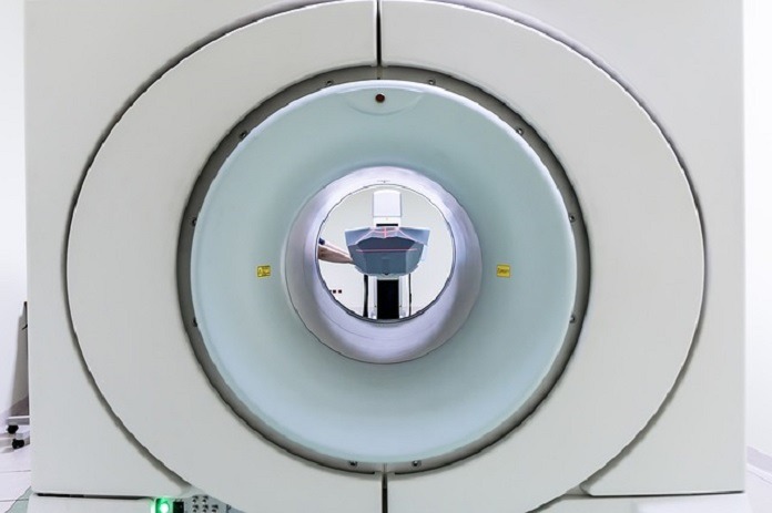
MRI and CT scans are both types of medical imaging used to diagnose many different medical conditions – what’s the difference between the two, and what are they used for?
What is an MRI and How Does it Work?
MRI, or magnetic resonance imaging, is a scanning procedure that generates detailed images of the body- ranging from cross-sections of organs to tissues. Using a combination of radio waves and magnetic fields, MRI tests can very accurately diagnose diseases, strokes, tumors, and more. In essence, MRI is able to accomplish this through a rapid process of reorienting certain subatomic particles that are present in the body’s water molecules.
Overview of an MRI Appointment
For a piece of equipment that can provide such complex information about a patient’s health, magnetic resonance imaging requires very little preparation before an appointment. It is important to note that because of the strong magnetic field generated inside, any metallic objects or accessories should not be worn during an MRI. An MRI may not be safe for people who have metallic implants.1
It is important that patients remain still while lying down during the scan, to prevent any interference with the production of MRI images.2 While the scanner is expected to generate loud banging noises, patients are encouraged to use headphones or earplugs to ease their experience.2
Impact of MRI on the Progression of Science
Along with its non-invasive design that does not cause the patient any pain or rarely experience any side effects, MRI has revolutionized medicine for doctors and researchers.
- MRI screening has a greater sensitivity for breast cancer detection in women than mammograms.3
- MRI has advantages over other diagnostic methods when investigating brain tumour segmentation.4
- Cine MRI, a special magnetic resonance imaging tool, is recognized as being the most accurate method of observing the function of ventricles in the heart.5
- MRI holds an increasingly crucial role in assessing patients with chronic liver disease, since the technology avoids the use of dangerous ionizing radiation.6
- Imaging produced by MRI is highly effective at categorizing the extent of male infertility.7
What is a CT Scan and How Does it Work?
Also referred to as a computerized tomography scan, a CT scan uses x-rays to produce intricate cross-sectional images of various parts of the body, as small as blood vessels. Merging a series of images produced from various angles, a CT scan functions exceptionally to examine and diagnose patients with internal injuries, tumors, diseases and more.
Concerns about CT Scans and Cancer
A considerable difference between an MRI and a CT scan is that CT scans rely on high frequency x-rays. Since this potentially harmful ionizing radiation is used, many scientific articles published over the course of recent years have raised concerns that CT scans may cause cancer.8
Although it is known that CT scans subject patients to higher doses of radiation in comparison to standard x-rays, studies have shown that these levels are still 10 to 100 times below the threshold that is expected to cause cancer.8 For this reason, the risk of cancer remains very low for only one CT scan. However, the risk is amplified following multiple scans.
Attempts to minimize these radiation risks to date have been complex, and will require great innovation, as well as time investments- especially from pediatric radiologists who are very concerned.9
Overview of a CT Scan Appointment
Patients entering a CT scan must remove any metal objects and ensure that they remain still during the examination, while x-rays are being taken from different angles. Once this data has been translated by computer software, cross-sectional images of particular areas of the body are produced.10
A significant difference between an MRI and a CT scan is the invasiveness of the procedure. Under certain circumstances, CT scans can be considerably more invasive and may require fasting prior to the examination. This is the case when a doctor recommends that a special dye referred to as contrast material must be injected, drank, or administered via the patient’s rectum, in order for their internal structures to be scanned more clearly.10
Renowned Uses of CT Scans in Medical Sciences
Similar to an MRI in scope, imaging from CT scans provides medical professionals with a range of life-improving applications.
- CT scans can accurately access incident spine and hip fractures for men and women over the age of 65.11
- Advancements in CT technology have played a crucial role for inspecting internal bleeding following torso trauma.12
- Researchers have used CT scans to facilitate the diagnosis and differentiation of rare intracranial tumours.13
- The fusion of images taken from real-time ultrasonography and CT has been successful at guiding needles into joints for sensitive injections.14
- CT scans have effectively been used to construct three-dimensional images of the distribution of human blood vessels.15
How Do These Imaging Methods Compare in Contemporary Medicine?
Although CT has long been the standard for diagnosing acute strokes, studies have revealed that this method no longer holds justifiable accuracy.16 Instead, research extensively comparing the differences between MRI and CT scan imaging has concluded that MRI is more effective for emergency diagnoses of acute strokes in a typical patient sample.16
Further reinforcing the benefits of using MRI, another study has concluded that magnetic resonance imaging is more accurate than CT when detecting chronic bleeding within brain tissue.17
While MRI is better at detecting certain medical irregularities that CT scans are incapable of seeing, that does not suggest that there are no other advantages of CT. For example, research has demonstrated that CT is superior to MRI in the diagnosis of certain mediastinal tumors that may arise in the chest.18
Therefore, while technical differences between MRIs and CT scans exist, both technologies are able to capture strikingly detailed images within the human body.
References
- Dempsey, M. F., Condon, B., & Hadley, D. M. (2002). MRI safety review. Seminars in ultrasound, CT, and MR, 23(5), 392–401. https://doi.org/10.1016/s0887-2171(02)90010-7
- Pullen R. L., Jr (2008). Preparing a patient for magnetic resonance imaging. Nursing, 38(10), 22. https://doi.org/10.1097/01.NURSE.0000337219.03593.e7
- Morrow, M., Waters, J., & Morris, E. (2011). MRI for breast cancer screening, diagnosis, and treatment. Lancet (London, England), 378(9805), 1804–1811. https://doi.org/10.1016/S0140-6736(11)61350-0
- Gordillo, N., Montseny, E., & Sobrevilla, P. (2013). State of the art survey on MRI brain tumor segmentation. Magnetic resonance imaging, 31(8), 1426–1438. https://doi.org/10.1016/j.mri.2013.05.002
- Ishida, M., Kato, S., & Sakuma, H. (2009). Cardiac MRI in ischemic heart disease. Circulation journal : official journal of the Japanese Circulation Society, 73(9), 1577–1588. https://doi.org/10.1253/circj.cj-09-0524
- Taouli, B., Ehman, R. L., & Reeder, S. B. (2009). Advanced MRI methods for assessment of chronic liver disease. AJR. American journal of roentgenology, 193(1), 14–27. https://doi.org/10.2214/AJR.09.2601
- Ammar, T., Sidhu, P. S., & Wilkins, C. J. (2012). Male infertility: the role of imaging in diagnosis and management. The British journal of radiology, 85 Spec No 1(Spec Iss 1), S59–S68. https://doi.org/10.1259/bjr/31818161
- McCollough, C. H., Bushberg, J. T., Fletcher, J. G., & Eckel, L. J. (2015). Answers to Common Questions About the Use and Safety of CT Scans. Mayo Clinic proceedings, 90(10), 1380–1392. https://doi.org/10.1016/j.mayocp.2015.07.011
- Rice, H. E., Frush, D. P., Farmer, D., Waldhausen, J. H., & APSA Education Committee (2007). Review of radiation risks from computed tomography: essentials for the pediatric surgeon. Journal of pediatric surgery, 42(4), 603–607. https://doi.org/10.1016/j.jpedsurg.2006.12.009
- Mayo Foundation for Medical Education and Research. (2020, February 28). CT Scan. Mayo Clinic. https://www.mayoclinic.org/tests-procedures/ct-scan/about/pac-20393675.
- Kopperdahl, D. L., Aspelund, T., Hoffmann, P. F., Sigurdsson, S., Siggeirsdottir, K., Harris, T. B., Gudnason, V., & Keaveny, T. M. (2014). Assessment of incident spine and hip fractures in women and men using finite element analysis of CT scans. Journal of bone and mineral research : the official journal of the American Society for Bone and Mineral Research, 29(3), 570–580. https://doi.org/10.1002/jbmr.2069
- Ahmed, N., Kassavin, D., Kuo, Y. H., & Biswal, R. (2013). Sensitivity and specificity of CT scan and angiogram for ongoing internal bleeding following torso trauma. Emergency medicine journal : EMJ, 30(3), e14. https://doi.org/10.1136/emermed-2011-200376
- Servo, A., Jääskeläinen, J., Wahlström, T., & Haltia, M. (1985). Diagnosis of intracranial haemangiopericytomas with angiography and CT scanning. Neuroradiology, 27(1), 38–43. https://doi.org/10.1007/BF00342515
- Klauser, A. S., De Zordo, T., Feuchtner, G. M., Djedovic, G., Weiler, R. B., Faschingbauer, R., Schirmer, M., & Moriggl, B. (2010). Fusion of real-time US with CT images to guide sacroiliac joint injection in vitro and in vivo. Radiology, 256(2), 547–553. https://doi.org/10.1148/radiol.10090968
- Chen, S. H., Chen, M. M., Xu, D. C., He, H., Peng, T. H., Tan, J. G., & Xiang, Y. Y. (2011). Anatomical study to the vessels of the lower limb by using CT scan and 3D reconstructions of the injected material. Surgical and radiologic anatomy : SRA, 33(1), 45–51. https://doi.org/10.1007/s00276-010-0702-9
- Chalela, J. A., Kidwell, C. S., Nentwich, L. M., Luby, M., Butman, J. A., Demchuk, A. M., Hill, M. D., Patronas, N., Latour, L., & Warach, S. (2007). Magnetic resonance imaging and computed tomography in emergency assessment of patients with suspected acute stroke: a prospective comparison. Lancet (London, England), 369(9558), 293–298. https://doi.org/10.1016/S0140-6736(07)60151-2
- Kidwell, C. S., Chalela, J. A., Saver, J. L., Starkman, S., Hill, M. D., Demchuk, A. M., Butman, J. A., Patronas, N., Alger, J. R., Latour, L. L., Luby, M. L., Baird, A. E., Leary, M. C., Tremwel, M., Ovbiagele, B., Fredieu, A., Suzuki, S., Villablanca, J. P., Davis, S., Dunn, B., … Warach, S. (2004). Comparison of MRI and CT for detection of acute intracerebral hemorrhage. JAMA, 292(15), 1823–1830. https://doi.org/10.1001/jama.292.15.1823
- Tomiyama, N., Honda, O., Tsubamoto, M., Inoue, A., Sumikawa, H., Kuriyama, K., Kusumoto, M., Johkoh, T., & Nakamura, H. (2009). Anterior mediastinal tumors: diagnostic accuracy of CT and MRI. European journal of radiology, 69(2), 280–288. https://doi.org/10.1016/j.ejrad.2007.10.002
- Image by Michal Jarmoluk from Pixabay


