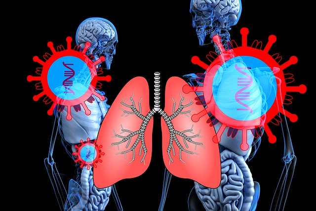
Researchers studied the imaging features of COVID-19 respiratory disease and investigated how they are similar to the features seen in SARS and MERS.
The novel coronavirus, COVID-19 is a highly infectious respiratory disease that first originated from Wuhan City, China and has been spreading globally. COVID-19 comes from the same family as severe acute respiratory syndrome (SARS) and Middle East respiratory syndrome (MERS). Similar to SARS and MERS, COVID-19 respiratory disease is diagnosed by manifestations such as fever, cough, malaise, and breathing difficulty.
Researchers reviewed the imaging from patients infected with SARS and MERS and investigated whether there were any similar changes in chest images that occur in patients with COVID-19.
The radiological investigations were reported in the American Journal of Roentgenology.
Chest images are an integral part of diagnosing and monitoring the progress of patients with respiratory illness. The chest images of acute and chronic stages of SARS and MERS are non-specific. About 80% of patients with SARS will have abnormal chest imaging. SARS imaging will display a unilateral, focal disease, with peripheral airspace opacity and a follow-up imaging will display mostly multifocal consolidation in one or both lungs. CT scan will display consolidation and patchy areas of ground-glass. Likewise, about 83% of patients with MERS will have abnormal chest imaging. MERS patients mostly exhibit multifocal airspace opacities in the lower lung areas on an x-ray or CT scan. Long-term follow-up imaging from a SARS infection will show transient reticular opacities and air trapping areas, rarely exhibiting fibrosis. With MERS, long-term follow-up imaging will show transient reticular opacities and about a third will have fibrosis.
COVID-19 imaging is also non-specific. With COVID-19 respiratory disease, the first imaging report of 40 out of 41 patients showed the involvement of both lungs in the first CT scan, displaying ground-glass pattern in non-ICU patients and consolidation in ICU patients. It was confirmed that 86% of patients had both lungs affected. 57% of cases had multifocal ground-glass opacities and 29% had consolidation.
Although the imaging results of COVID-19 respiratory disease are similar to that of SARS and MERS, it has been observed that COVID-19 imaging involves both lungs while SARS and MERS are usually unilateral. It is important to note that an early CT scan with normal results does not exclude the infection.
One of the authors, Melina Hosseiny concluded, “Early evidence suggests that initial chest imaging will show abnormality in at least 85% of patients, with 75% of patients having bilateral lung involvement initially that most often manifests as subpleural and peripheral areas of ground-glass opacity and consolidation.”
She also added, “older age and progressive consolidation” may imply an overall poorer prognosis.” “To our knowledge,” Hosseiny et al. continued, “pleural effusion, cavitation, pulmonary nodules, and lymphadenopathy have not been reported in patients with COVID-19.“
References:
1. Hosseiny, M., Kooraki, S., Gholamrezanezhad, A., Reddy, S. and Myers, L. (2020). Radiology Perspective of Coronavirus Disease 2019 (COVID-19): Lessons From Severe Acute Respiratory Syndrome and Middle East Respiratory Syndrome. American Journal of Roentgenology, pp.1-5.
2. EurekAlert!. (2020). AJR: Novel coronavirus (COVID-19) imaging features overlap with SARS and MERS. [online] Available at: https://www.eurekalert.org/pub_releases/2020-02/arrs-anc022820.php [Accessed 3 Mar. 2020].
Image by Gerd Altmann from Pixabay


