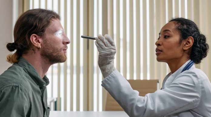
Moorfields Eye Hospital London experts, led by Pearse Keane, have developed an Artificial Intelligence tool to provide a fast and reliable diagnosis for patients with eye-related diseases.
In recent years, there has been a rapid push to integrate artificial intelligence (AI) into healthcare systems. Artificial Intelligence (AI) serves to train a machine to make precise, swift, and efficient decisions. In medical imaging for example, AI plays a pivotal role by evaluating graphics such as an X-ray or CT scans to determine whether a patient has an underlying medical condition.1
A New Artificial Intelligence Tool
A recent study published in Nature presented a novel AI model, RETFound.2 In a press release Dr. Pearse Keane, a consultant ophthalmologist from the Moorfields Eye Hospital NHS Foundation Trust explains,
“This is another big step towards using AI to reinvent the eye examination for the 21st century, both in the UK and globally. We show several exemplar conditions where RETFound can be used, but it has the potential to be developed further for hundreds of other sight-threatening eye diseases that we haven’t yet explored.
“If the UK can combine high quality clinical data from the NHS, with top computer science expertise from its universities, it has the true potential to be a world leader in AI-enabled healthcare. We believe that our work provides a template for how this can be done.”3
An AI tool that can accurately and consistently diagnose retinal disease could revolutionize the ability of doctors to preserve peoples sight in regions with limited access to ophthalmologists.
The retina is the light detecting sensory area at the back of the eye. It uses densely packed photoreceptors to capture light and convert it to a signal that is sent to the brain.4 Many health conditions can contribute to damage to this delicate organ. Retinopathies (disease of the retina) can reduce the amount of working light-receptors in the eye, eventually leading to vision loss. Early detection allows doctors to treat the cause of the damage, slowing or stopping it in time to save your sight. This is why your optician is so insistent that you get your eyes dilated!
The First AI Foundation Model Developed for Disease Detection
A foundation model is an AI model that can learn from huge datasets (such as retinal images) to generate many different downstream tasks and outputs. RETFound is one of the first AI foundation model’s created in healthcare for disease detection.
In contrast to foundation models, developers designed early AI models to perform very specific and limited tasks. Foundation models like RETFound are distinct from previous AI models, a versitile tool, they can adapt to a range of jobs and outputs .5 A foundation model that most of us know is ChatGPT.6
Exceptional Performance and Efficiency
The team supplied RETFound roughly 1,640,612 retinal scans from patients examined at Moorsfield Eye Hospital, between 2000 and 2022. They then instructed the RETFound programme to use Self-Supervised Learning to practice recognizing signs of retina disease.
The researchers then acquired publicly available retina scans from international databases and deployed RETfound to perform a diagnosis. In parallel, a team of medical experts, retina specialists, ophthalmologists and senior retina specialists, annotated each image with a diagnosis and a grade of severity. Where experts encountered conflicting opinions, a panel of five senior retina specialists reviewed and resolved the disagreement. They then compared the results of the analyses by RETfound to the diagnoses by the doctors, and scored for how well they agreed.
RETFound successfully diagnosed ocular diseases such as glaucoma and diabetic retinopathy. Encouragingly, it outperformed existing AI tools such as SSL-ImageNet and SSL-Retinal.
Streamlined Technology
Development of AI models requires an enormous effort. A small army of ophthalmologists were gathered to review and to label images of retinas from patient files. The developers fed annotated pictures into the algorithm to teach the AI what a normal retina looked like and how a diseased retina looked.
A remarkable aspect of RETFound was its efficiency. RETFound required only 10% of the manually added labels required by other models, reducing training time by up to 80% and reducing manual annotation work by doctors. Not only did this new tool save time, but it also allowed more diverse training input images. This will allow effective detection of retinal diseases across varied populations.
RETFound sets itself apart from current tools
Besides discerning whether a retina in an image looks healthy or not, RETFound can distinguish between a picture of a retina damaged due to heart failure, and a retina characteristic of Parkinson’s disease, stroke or heart attack amongst others. This strength allows the AI tool to tell the ophthalmologist not only that a person has an unhealthy retina, but also that they may have had a stroke and should be referred to a neurologist.
This ability to perform multiple tasks with different outputs is a step forward for the technology. For example, ImageNet, a competing AI model, was developed and pre-trained to recognize images such as an image of a dog, the street, buildings.7 However, it failed to adapt to new datasets after undergoing extensive pre-training on other data sources. SL-ImageNet uses supervised learning to pre-train the model, which limits its ability to discern only low-level features such as lines and curves.2 In contrast, RETFound is expected to expand and develop further into detecting other vision-related diseases, a huge advantage to clinicians.
Taking The Long View
The authors of the study acknowledge that despite its remarkable performance, RETFound faced obstacles when tested on cohorts with differing demographics, particularly when predicting systemic diseases.
A challenge for developers is the enormous medical data required to teach this model and the multiple computational resources required to develop this model.
They also need to train RETfound on more diverse populations. Currently RETFound is optimized for people living in the UK and might be more familiar with morphological quirks in the retina that are peculiar to North West European populations than others.
A Promising Future
RETFound is the first medical foundation model that can perform a wide range of downstream tasks. This innovation promises to enhance timely diagnosis of ocular and systemic diseases. As medicine continues its rapid advancement, the integration of AI promises significant benefits for both patients and medical professionals.
References
1. Panayides, A. S., Amini, A., Filipovic, N. D.,et al. AI in medical imaging informatics: current challenges and future directions. IEEE journal of biomedical and health informatics. 2020 24 (7), 1837-1857.;doi: 10.1109/JBHI.2020.2991043.
2.Zhou, Y., Chia, M. A., Wagner, S. K., Ayhan, M. S. et al. A foundation model for generalizable disease detection from retinal images. Nature. 2023; 1-8. doi.org/10.1038/s41586-023-06555-x
3.World-first AI foundation model for eye care to supercharge global efforts to prevent blindness. UCL Media Centre. Published 13th September 2023, Accessed 20th September 2023.https://www.ucl.ac.uk/news/2023/sep/world-first-ai-foundation-model-eye-care-supercharge-global-efforts-prevent-blindness
4. Mahabadi N, Al Khalili Y. Neuroanatomy, Retina. Updated 2023 Aug 8. In: StatPearls [Internet]. Treasure Island (FL): StatPearls Publishing; 2023 Jan-. Available from: https://www.ncbi.nlm.nih.gov/books/NBK545310/
5. Lutkevich, B. (2023, August 8). Foundation models explained: Everything you need to know. WhatIs.com. https://www.techtarget.com/whatis/feature/Foundation-models-explained-Everything-you-need-to-know
Zhou, C., Li, Q., Li, C. et al. A comprehensive survey on pretrained foundation models: A history from bert to chatgpt. arXiv preprint arXiv:2302.09419.
Guo, Y., Liu, Y., Bakker, E. M., Guo, Y., & Lew, M. S. CNN-RNN: a large-scale hierarchical image classification framework. Multimedia tools and applications. 2018; 77(8), 10251-10271.2. DOI:10.1007/s11042-017-5443-x


